| |
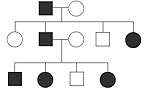
Животные модели для изучения генетически обусловленных глазных болезней
Списком представлены модели животных (в основном мыши и крысы), на которых разрабатываются способы лечения глазных заболеваний.
264 - D0213. Identification of a novel genetic model for retinal neovascularization
Allocca, Mariacarmela 1 ; Leehy, Barrett D. 1 ; White, Joseph A. 1 ; Kim, Leo A. 1 ; Saint-Geniez, Magali 1 ; Escher, Pascal 2, 3 ; Eliott, Dean 1 ; Pierce, Eric A. 1 ; DeAngelis, Margaret M. 4 ; Haider, Neena B. 1
1. Massachusetts Eye and Ear/Schepens Eye Research Institute, Department of Ophthalmology, Harvard Medical school , Boston, MA, United States.
2. IRO-Institute for Research in Ophthalmology, Sion , Switzerland.
3. Department of Ophthalmology, University of Lausanne, Lausanne, Switzerland.
4. Moran Eye Center, University of Utah, Salt lake City, UT, United States.
The Nrv1 mouse exhibits abnormal production of de novo blood vessels in the retina that innervate into the vitreous. The blood vessels in the retina are disorganized and clumped. A disorganized astrocytes network underlines the abnormal vascular topography. The abnormal blood vessels into the vitreous originate from retinal vasculature and are preceded by the uncommon presence of astrocytes into the vitreous. The overall data suggests misguidance defects due to the astrocytes. Linkage analyses mapped the vascular disorder to a 1Mb region on the chromosome 9. We are currently sequencing this region in order to identify the mutation and the causative gene.
265 - D0214. Survey of Common Genetic Eye Diseases in Mouse Strains
Bo Chang ; Ron Hurd; Jieping Wang; Patsy M. Nishina
1. The Jackson Laboratory, Bar Harbor, ME, United States.
266 - D0215. Up-regulation Of Growth Hormone Releasing Hormone (GHRH) Receptors In The Ischemic Retina
Patel, Sagar 1 ; Lamoke, Folami 1, 2 ; Stampley, Chaunte E. 3 ; Baban, Babak 4 ; Romero, Maritza 2 ; Lucas, Rudolf 5, 2 ; Block, Norman L. 6, 7 ; Schally, Andrew 6, 7 ; Bartoli, Manuela 1, 2
1. Department of Ophthalmology, Georgia Health Sciences University, Augusta, GA, United States.
2. Department of Pharmacology and Toxicology, Georgia Health Sciences University, Augusta, GA, United States.
3. Rollins School of Public Health, Emory University, Atlanta, GA, United States.
4. Department of Oral Biology, Georgia Health Sciences University, Augusta, GA, United States.
5. Vascular Biology Center, Georgia Health Sciences University, Augusta, GA, United States.
6. Veteran Affairs Medical Center and South Florida Veterans Affairs Foundation for Research and Education, Miami, FL, United States.
7. Miller School of Medicine, University of Miami, Miami, FL, United States.
Growth hormone releasing hormone (GHRH) is a hypothalamic hormone involved in the regulation of growth hormone release from the pituitary gland. Recent evidence suggests that GHRH expression and activity has extra-pituitary effects. Here we have studied the localization and expression of GHRH receptors (GHRH-R) in normal and ischemic retina.
269 - D0218. Downregulation of aldehyde detoxification enzymes in the diabetic retina
Rosie Mcdowell ; Timothy M. Curtis; Graham McGeown
1. Centre for Vision and Vascular Science, Queens University Belfast, Belfast, United Kingdom.
RNA from the retinas of Sprague Dawley rats was isolated to determine the expression of aldehyde detoxification genes by RT-PCR. For immunohistochemistry, eyes were subject to fixation in 4% PFA, cryo-sectioning and then labelling with anti-bodies against ALDH1a1, ALDH2, AKR1B1 and AKR7a2. In separate experiments, male Sprague-Dawley rats were rendered diabetic by a single injection of streptozotocin (65mg/kg) and eye tissue was harvested after 3 months disease duration. RNA was isolated from the retinas of six diabetic animals and six age matched sham controls and then processed for RT-PCR. All animal care and experimental procedures strictly conformed to the ARVO Statement for the use of Animals in Ophthalmic and Vision Research. RT-PCR indicated ALDH1a1, 2, 3a1, 3a2, 9, 16, 18a1 AKR1b1 and 7a2 were expressed in the rat retina. Immunohistochemistry experiments revealed that ALDH1a1, 2, AKR1B1 and 7a2 are principally localised to retinal Muller cells. The comparison of mRNA transcripts between control and diabetic animals indicated that ALDH1a1 7a2 and 3a2 were significantly down-regulated in whole retinal extracts of diabetic animals, whilst polyamine metabolic enzyme levels were unchanged.
Down-regulation of detoxifying enzymes in retinal Muller cells may represent a major mechanism contributing to the selective accumulation of FDP-lysine adducts in these cells during diabetes.
270 - D0219. TXNIP regulates neurotrophic factor expression and neuronal injury in early diabetic retinopathy
Singh, Lalit P. 1, 2 ; Devi, Takhellambam S. 1
1. Anatomy and Cell Biology , Wayne State Univ Sch of Med, Detroit, MI, United States.
2. Ophthalmology, Wayne State Univ Sch of Med, Detroit, MI, United States.
recently published that the pro-oxidant thioredoxin interacting protein (TXNIP) is significantly up-regulated in the diabetic retina in vivo and mediates cellular oxidative stress and retinal inflammation. The purpose of this study is to investigate the role TXNIP plays in neurotrophic factor expression and neuronal injury in early diabetic retinopathy (DR).
Streptozotocin (STZ)-induced diabetic and age-matched normal rats were maintained for 8 weeks. Subsequently, retinas were harvested and analyzed for gene expression by real time quantitative PCR and immunohistochemistry. To knock down TXNIP, scrambled RNA and siTXNIP were injected intravitreally in diabetic rat retinas.
TXNIP, iNOS, radial GFAP (Muller cell (MC) activation) and protein s-nitrosylation (SNO) are increased in diabetic rat retinas when compared with normal rats. These results suggest that TXNIP up-regulation correlates with retinal oxidative/nitrosative (ROS/RNS) stress and MC activation. In addition, we found that the mRNA level of brain-derived neurotrophic factor (BDNF) is not altered; however, the expression of its high affinity receptor TrkB and that of glia derived neurotrophic factor (GDNF) are significantly increased. Conversely, the mRNA levels of dendritic spine protein synaptopodin and tyrosine hydroxylase (TH), a marker for dopaminergic inter-neurons, are down-regulated in DR indicating neuronal injury. Finally, knock down of TXNIP by siRNA reduces several molecular abnormalities of early DR.
TXNIP expression is critical in the initiation and development of early DR. Therefore, TXNIP represents a novel therapeutic target to ameliorate blinding ocular complications of diabetes.
273 - D0222. Profiling miRNAs in a hyperglycemic and hypoinsulinemic Ins2Akita mouse model
Giocanti-Auregan, Audrey 1, 2 ; Ahn, Lisa 1 ; Sancho-Pelluz, Javier 3, 4 ; Tsang, Stephen H. 3 ; Kiss, Szilard 1 ; Rosenblatt, Mark 1 ; Tuschl, Thomas 5 ; Pena, John T. 1 ; D'Amico, Donald J. 1
1. Ophthalmology, Weill Cornell Medical College, New York, NY, United States.
2. Ophthalmology, Avicenne hospital, Bobigny, France.
3. Bernard & Shirlee Brown Glaucoma Laboratory. Department of Ophthalmology, Columbia University, New York, NY, United States.
4. Facultad de Medicina, Universidad Catolica San Vicente Martir, Valencia, Spain.
5. Howard Hughes Medical Institute , The Rockefeller University, New York, NY, United States.
275 - D0224. Differential Distribution of Optic Nerve Lipids in the DBA/2J Mouse Model of Glaucoma
Sakrikar, Dhananjay 1, 2 ; Anderson, David M. 1, 2 ; Spraggins, Jeffrey M. 1 ; Lambert, Wendi S. 3 ; Schey, Kevin L. 1, 2 ; Calkins, David J. 3
1. Mass Spectrometry Research Center, Vanderbilt University, Nashville, TN, United States.
2. Biochemistry, Vanderbilt University, Nashville, TN, United States.
3. Vanderbilt Eye Institute, Vanderbilt University, Nashville, TN, United States.
DBA расшифровывается как
Dilute Brown Non-Agouti - светлокоричневый окрас. 3, 6, and 9-month-old DBA/2J mice were used in this study. Whole eyes including optic nerve were removed and rapidly frozen in carboxymethyl cellulose. A cryostat was used to collect 12 µ thick tissue sections that were then thaw mounted onto a gold-coated plate. The sections were desiccated and washed with 100 mM ammonium acetate before sublimating the MALDI matrix, 1,5-Diaminonaphthalene. Imaging was performed using a Bruker UltrafleXtreme II and lipid identification was performed using a 9.4T Bruker Apex Qe FT-ICR mass spectrometer. FlexImaging software was used to generate images. MALDI imaging was performed in both positive and negative ion mode to identify different lipid species from the optic nerve. MALDI-IMS produced signals for a number of phosphocholine and ceramide species of lipids. As expected, a few lipids maintained similar spatial distributions across the age groups in DBA/2J mice. However, lipid species such as LPC (18:0/0:0), PC (18:1/18:0), PC (18:0/20:4), PC (18:0/22:6) in positive ion mode and sulphoHex-Cer (d18:1/18:0), sulphoHex-Cer (d18:1/22:0), sulphoHex-Cer (d18:1/22:0(2OH)) in negative ion mode showed differences in relative abundance and/or spatial distribution in the disease model with age.
Here we report alterations in relative abundance and differential localization of multiple lipid species in the DBA/2J mouse model of glaucoma with age. Some of these lipid species may be involved in cell signaling. Understanding the changes associated with lipid distributions in optic nerve tissue will further elucidate the underlying mechanism and signaling changes associated with glaucoma.
Мутация генов Gpnmb и Tyrp1 у мышей вызывает изменения радужной оболочки, дисперсию пигмента и как следствие - повышение внутриглазного давления. (http://www.biomedcentral.com/1471-2156/8/45). Подробное описание линии мышей DBA здесь: http://jaxmice.jax.org/strain/000671.html
A transcriptomic Approach to Refine the Primary Locus of a Canine Oligogenic Cone-Rod Dystrophy
Das, Gautami 1 ; Swaroop, Anand 2 ; Brooks, Matthew 2 ; Aguirre, Gustavo D. 1 ; Miyadera, Keiko 1
1. School of Vet Medicine, Univ of Pennsylvania, Philadelphia, PA, United States.
2. National Eye Institute, NIH, Bethesda, MD, United States.
Cone-rod dystrophies (CRDs) cause cone dysfunction/loss prior to rod death, and eventually lead to blindness. A research colony was used to map a canine CRD (cord1) to a 14Mb interval on CFA15, and a homozygous exonic 44 bp insertion (ins/ins) resulting in a premature stop was identified in a positional candidate gene RPGRIP1. The interval was subsequently narrowed down to 1.74Mb still containing RPGRIP1. We have since identified a modifier locus, establishing an oligogenic model where a modifier controls the age of clinical onset of a cone-led retinal degeneration caused by homozygosity at the primary locus linked to RPGRIP1. In a recently reestablished research colony, however, some RPGRIP1ins/ins dogs still retain cone-ERG, while others develop no or severely diminished responses. Our aim was to refine the primary cord1 locus underlying cone-ERG loss.
RNA-seq was performed by high-throughput sequencing of retinal cDNA libraries from these dogs using Illumina GAIIx, followed by assembly and analysis of the sequence reads. Differential expression of annotated and unannotated transcripts/genes was analyzed across the interval. Alternatively spliced isoforms of RPGRIP1 and other genes in the interval were assessed qualitatively and quantitatively. Based on sequence variants identified within the interval, haplotypes were constructed.
Of the 24 annotated genes across the 1.74Mb cord1 primary locus critical interval, 23 showed retinal expression. Neither these genes nor their isoforms showed significant expressional difference between the two phenotypic groups. Additional reads were mapped within introns and immediate flanking regions of annotated genes. No new retinally expressed genes were identified within the interval. SNP-based haplotype analysis across this interval did not reveal any difference between the two studied groups.
Expression of known retinally expressed genes/transcripts across the cord1 primary locus was comparable in normal and cone loss RPGRIP1ins/ins dogs. The shared haplotype spanning the primary locus among all RPGRIP1ins/ins dogs regardless of the phenotype indicated the involvement of factor(s) beyond the critical interval in cone function loss.
Sanchez, Maria C. 1 ; Lorenc, Valeria E. 1 ; Jose, Luna D. 2 ; Chiabrando, Gustavo A. 1
1. CIBICI-Dpto de Bioquimica Clinica, Fac de Ciencias Quimicas UNC, Cordoba, Argentina.
2. Clinica de Ojos Romagosa-Funadacion Ver, Cordoba, Argentina.
Neonatal C57BL/6J mice were subjected to 75% oxygen from postnatal day 7 (P7) to P12 and then returned to room air for five days. Control mice were exposed to room air from birth until P17. Retinal blood vessel patterns were visualized by GSA labeling. Retinas from animals with and without OIR treatment were analyzed for IGF-1R expression and tissue distribution at selected time points (P3, P12, P15, P18, P21 and P27). The IGF-1R localization was examined using cell-type specific markers (GFAP, GS, Brn3a, PKC alpha and calbindin among other) by immunofluorescence and confocal microscopy. In order to identify apoptotic cells in the same retinas, TUNEL assay (Roche) was used.
In retinas without OIR we first observed IGF-1R expression in endothelial cells which was confirmed by using the GSA. This receptor was also expressed at level of ganglion cells as well as in the end feet of Muller glial cells. In addition, we visualized staining for IGF-1R in inner and outer nuclear layers. When the IGF-1R expression in the OIR model was analyzed, we observed a change in the distribution due to an alteration in the structure of the neural retina. Considering that this model produces Muller glial cells activation and retinal cell death, then the expression of GFAP was analyzed. Increased GFAP expression was detected in Muller cells in OIR retinas from P15. Finally, in agreement with GFAP expression, TUNEL possitive cells in the retinas were also observed at P15.
The IGF-1R distribution and localization in retinas were modified after OIR treatment coinciding with the onset of GFAP expression and the retinal cell death.
Photoreceptors are a main generator of superoxide in retinas of diabetic mice
Kern, Timothy S. 1, 2 ; Du, Yunpeng 1 ; Palczewski, Krzysztof 3 ; Berkowitz, Bruce A. 4
1. Medicine, Case Western Reserve University, Cleveland, OH, United States.
2. Stokes Veterans Administration Hospital, Cleveland, OH, United States.
3. Pharmacology, Case Western Reserve University, Cleveland, OH, United States.
4. Anatomy/Cell Biol, Wayne State Univ Sch of Med, Detroit, MI, United States.
rior work by us and others has provided strong evidence that oxidative stress and inflammatory processes play important roles in the development of the vascular lesions of early diabetic retinopathy. Oxidative stress is known to regulate expression of pro-inflammatory proteins, thus making it important to understand the source of the oxidative stress. We have investigated the contribution of photoreceptors to the retinal oxidative stress and induction of proinflammatory ICAM-1 in diabetes.
Retinal oxidative stress was assessed histologically using dichlorofluorescein staining, and quantitatively using luciginen luminescence in C57Bl/6J mice (nondiabetic and diabetic for 2 mos). Two models that caused degeneration of photoreceptors (a genetic model (rhodopsin knockout) and a chemically-induced degeneration of photoreceptors (iodoacetic acid)) were studied after 2 mos diabetes.
In the diabetic mice, dichlorofluorescein stain indicated that the majority of oxidative stress was localized in the photoreceptor layer. Both models of photoreceptor degeneration resulted in essentially total obliteration of photoreceptors. The diabetes-induced increase in superoxide observed in wildtype C57Bl/6J mice was significantly inhibited in both of the diabetic models lacking photoreceptors. The diabetes-induced induction of ICAM-1 (used as a marker of inflammation) was significantly inhibited in diabetic animals missing photoreceptors (rhodopsin knockout).
These findings demonstrate a critical role of photoreceptors in the diabetes-induced oxidative stress in the retina, and presumably in the pathogenesis of other lesions of diabetic retinopathy.
An Xbp1-GFP-rhodopsin reporter transgene for assessing ER stress in Xenopus laevis models of retinitis pigmentosa
Tam, Beatrice M. 1 ; Lin, Jonathan H. 2 ; Moritz, Orson L. 1
1. Ophthalmology and Visual Sciences, University of British Columbia, Vancouver, BC, Canada.
2. Pathology, UCSD School of Medicine, La Jolla, CA, United States.
We have previously demonstrated that in transgenic X. laevis, P23H rhodopsin is significantly retained in the ER, and that light exposure exacerbates this ER retention and promotes retinal degeneration. Here we examine whether accumulation of the mutant protein induces ER stress in vivo by monitoring the splicing of an Xbp1-GFP reporter construct.
Methods: The DNA binding domain of murine Xbp1 was deleted and an out of frame cDNA encoding a GFP-rhodopsin fusion protein inserted downstream of the unconventional splice site. Upon ER stress, splicing of the mRNA produces an in-frame Xbp1-GFP-rhodopsin (XGR) fusion protein. This engineered cDNA was used to generate transgenic X. laevis expressing the fusion protein under the control of the opsin promoter. An XGR frog was mated with a transgenic frog expressing bovine P23H rhodopsin (bRhoP23H). The resulting tadpoles were raised for 14 days in constant dark, which prevents retinal degeneration in this animal model. Half the tadpoles were then exposed to two 12:12 light:dark cycles and returned to the dark. The other half were maintained in constant dark. On day 21, all tadpoles were killed and their eyes used for dot blot analysis or for fluorescence microscopy.
Results: In our line of XGR animals, GFP fluorescence peaked shortly after the onset of rod opsin expression (~d5) and thereafter declined and was virtually undetectable by d21. Interestingly, XGR distributed to both inner and outer segment membranes. In contrast, a similar protein (GFP-RhoCT44) which does not include Xbp1, is almost exclusively delivered to the outer segment. In cryosections of eyes that were not expressing bRhoP23H, little or no GFP fluorescence was observed regardless of whether the animals had received light exposure or not. Similarly, dark reared bRhoP23H retinas expressed minimal XGR. In contrast, light exposed bRhoP23H retinas exhibited higher levels of fluorescence. Furthermore, nuclear XGR localization was observed in this group. As expected, retinas expressing bRhoP23H underwent retinal degeneration when exposed to light.
Conclusions: At d21, significant levels of Xbp-GFP-RhoCT44 were observed only in light exposed animals expressing bRhoP23H. This is consistent with a pathogenic role for light-induced P23H rhodopsin misfolding and ER stress in this model of retinitis pigmentosa.
Analysis of microRNA expression changes following explanation of bovine RPE
Redmond, T. Michael 1 ; Jaworski, Cynthia 1 ; Samuel, William 1 ; Kutty, R K. 1 ; Duncan, Todd 1 ; Parikh, Toral P. 1, 2
1. LRCMB Bldg 6 Rm 117A, National Eye Inst/NIH, Bethesda, MD, United States.
2. Howard Hughes Medical Institute, Bethesda, MD, United States.
An enduring challenge in studying retinal pigment epithelium (RPE) is the limited quantity of tissue available. Although cultured RPE cells are widely used, the phenotypes of cultured cells, with time, differ substantially from native tissue. For example, many cultured RPE cells do not express RPE65 protein, a distinguishing feature of RPE. To better understand this alteration in gene expression, in particular the loss of key visual cycle proteins, we asked whether the expression pattern of microRNAs (miRs) is altered after explantation, in concert with the changes in gene and protein expression.
Methods: RPE was harvested from freshly dissected bovine eyes and placed in tissue culture. RNA and protein were extracted by standard means from fresh tissue and from cells harvested after 4 or 8 weeks in culture. MiR expression was assessed using the miRCURY LNA miR Array (Exiqon Services). Quantified signals were normalized. Expression levels of selected miRs were validated by real time RT-PCR.
Results: Principal component analysis indicates that samples cluster according to their time in culture. Hierarchical clustering reveals the specific miRs comprising each cluster. Nineteen miRs were significantly different (p-value less than 0.05 with Bonferroni multiple testing) between native tissue and 4-week cultures; 78 differences were found between native tissue and 8-week cultures. There were 28 microRNAs that differed between the 4 and 8 week culture samples. Several miRs show evidence of a transient change of expression at 4 weeks, returning to levels similar to that of native tissue upon more prolonged culture time. Interestingly, the “sensory cluster” of miR-96, -182, and -183 all had diminished expression with culture, as did mir-204 and -211, reported to be characteristic of RPE. Real time RT-PCR of selected miRs corroborated the expression profiles. Expression of miR-21 and -184 rose markedly with growth in culture; sensory cluster miR levels fell.
Conclusions: The pattern of microRNA expression changes significantly when bovine RPE cells are explanted and grown in culture, suggesting that this class of regulatory molecules may be important in producing the altered culture phenotype and/or in maintaining the characteristics that define a functional in situ RPE cell. Analysis of potential gene targets showing increase or decrease will help us discriminate between miRs regulating these opposing trends.
Accumulated Increase of mtDNA Damage Contribute to Progressive Loss of RGC in a Rat Model of Glaucoma
Wu, Jihong 1 ; Zhang, Shenghai 1
1. Eye & ENT Hospital, Fudan University , Shanghai, China.
Wistar rat was used to induce glaucomatous model through episcleral vein cauterization (EVC). IOP elevation sustained for 6 weeks and we continued the experiment till 6 months when IOP had already returned to normal to mimic the clinical feature of glaucoma. RGC loss was detected by retrograde labeling of FG. Fresh RGCs was isolated with magnetic beads coated by CD 11b/c and Thy-1 antibodies. Mitochondria isolation, DNA and RNA isolation were carried out in order to detect mtDNA damage or mutations, mtDNA copies, expression of mtDNA repair enzymes, and mitochondrial function. Cultured astrocytes were given 30 mmHg to study the whether pressure induce mitochondria dysfunction and possible mechanisms.
Conditional mutant analysis of Dnmts in murine retinal ganglion cells
Enke, Raymond 1 ; Yang, Zhiyong 1 ; Hauswirth, William W. 2 ; Boye, Sanford L. 2 ; Chiodo, Vince 2 ; Zack, Donald J. 1 ; Merbs, Shannath L. 1
1. Ophthalmology, Johns Hopkins University, Baltimore, MD, United States.
2. Ophthalmology, University of Florida, Gainesville, FL, United States.
Epigenetic mechanisms may modulate the onset and progression of disease in the retina. As a prelude to studying the role of DNA methylation in glaucoma and optic nerve degeneration, we set out to determine the expression pattern of DNA methyltransferase (Dnmt) enzymes in mature murine retinal ganglion cells (RGCs). We also use conditional Dnmt mutant lines to demonstrate RGC survival following optic nerve crush.
Methods: Eyes from adult wt mice were fixed in 4% PFA, equilibrated in a sucrose gradient, and cryopreserved. Retinal cross sections were cut and used for immunohistochemical (IHC) analysis of Dnmts. Conditional Dnmt1, Dnmt3a, and Dnmt3b mutants were created by intravitreally injecting an adeno-associated virus serotype 2 (AAV2) encoding Cre recombinase into transgenic mouse lines harboring lox P sites in each respective allele. Optic nerve crush was performed in adult mice to induce RGC degeneration. For RGC viability analysis, eyecups were fixed in 4% PFA and used for whole mount IHC quantification of Brn3 and Tuj1. Corresponding optic nerve cross sections were also stained for phosphorylated neurofilament (pNF).
Results: IHC analysis of the adult mouse retina demonstrated that Dnmt1 and Dnmt3b are expressed in postmitotic RGCs while Dnmt3a expression is limited to amacrine cells in the ganglion cell layer (GCL) of the retina. Intravitreal delivery of AAV2-Cre results in efficient transduction of the mouse GCL cells and proves to be an effective technique for producing conditional Dnmt mutant mice. Dnmt1 mutant RGCs were partially protected from degeneration following optic nerve crush. Surviving Dnmt1 mutant RGCs also displayed more persistent expression of Brn3 following optic nerve crush compared to controls.
Conclusions: Expression of Dnmt1 and Dnmt3b in postmitotic RGCs suggests that DNA methylation may be involved in RGC homeostasis. Our current experiments further address this question by assaying the role of Dnmts in RGC survival following optic nerve injury. A conditional mutation in Dnmt1 imparts partial protection against RGC degeneration following optic nerve crush. This finding indicates that DNA methylation may be an important epigenetic signal in response to optic neuropathy. Ongoing analysis will determine if Dnmt3b conditional mutants also have improved RGC viability following optic nerve crush.
Adenosine triphosphate (ATP) Signaling Pathways Trigger Glial Activation in the Mouse Retina
Mac Nair, Caitlin E. 1, 2 ; Schlamp, Cassandra 2 ; Nickells, Robert W. 2
1. Cellular and Molecular Pathology Graduate Program, Univeristy of Wisconsin, Madison, WI, United States.
2. Ophthalmology and Visual Sciences, Univeristy of Wisconsin, Madison, WI, United States.
Optic nerve injury causes RGC death and activation of the retinal macroglia and microglia. Bax -deficient RGCs are resistant to this acute injury and display an attenuated glial activation response, indicating a relationship between cell death and glial activation. Several studies suggest that injured neurons release ATP as a distress signal, and we investigated the potential role of ATP in triggering glial activation, the consequence of which may lead to cytokine production that damages additional ganglion cells.
Methods: Wild type mice were intravitreally injected with 1?l of a 250?M ATP receptor agonist, either ATP?S or BzATP, and mRNA and protein levels of glial activation markers (GFAP for macroglia and AIF1 for microglia) were monitored by qPCR and immunofluorescence. RGC gene markers Thy1 , Nrn1 , and Sncg were also monitored by qPCR. Additional experiments will include Bax -deficient mice injected with either BzATP or ATP?S, and wild type mice treated with an ATP receptor antagonist, either oxATP or PPADS, to inhibit ATP signaling from crush injury. The effects on glial activation and RGC survival will be reported.
Results: Bax -deficient RGCs were completely resistant to optic nerve crush and displayed an attenuated macroglial and microglial activation response, suggesting that cell death is required for glial activation. A single intravitreal injection of BzATP, a specific P2X 7 receptor agonist, caused a significant spike in macroglial activation by 24 hours with prominent labeling in the Muller cells. By 48 hours macroglial activation returned to basal levels and remained unchanged by 72 hours. Contrary, macroglial activation by the broad spectrum P2X receptor agonist, ATP?S, showed a moderate increase at 24 hours above vehicle-injected eyes but continued to rise at 48 and 72 hours, with strong labeling of the Muller cells by 72 hours. A decline in RGC gene markers, indicative of RGC damage, was also apparent at 72 hours in ATP?S-injected eyes. Microglial activation was not greatly affected by either treatment.
Conclusions: These results indicate that ATP may contribute to macroglial activation during glaucoma. The sustained elevation of ATP?S over BzATP at 72 hours suggests that purinergic receptors in addition to P2X 7 may be involved in triggering activation and subsequent injury to RGCs.
Whirlin proteins localize at the outer limiting membrane and subapical region of zebrafish retina
Toro, Sabrina 1 ; Phillips, Jennifer B. 1 ; Westerfield, Monte 1
1. Institute of Neuroscience, University of Oregon, Eugene, OR, United States.
Mutations in DFNB31 cause Usher syndrome type 2D. DFNB31 encodes Whirlin (Whrn), a scaffold protein thought to mediate interactions with other Usher proteins. We previously reported the discovery of two dfnb31 genes in zebrafish, each with multiple splice variants. Here, we investigate the subcellular localizations and functions of WhrnA and WhrnB in the zebrafish retina.
Methods: Immunohistochemistry on cryosectioned tissues was visualized by confocal microscopy. Antibodies were generated to unique regions of WhrnA and WhrnB, and the third PDZ domain (Whrn_PDZ3) common to both proteins. Antibodies against Harmonin (ush1c), Usherin (ush2a), Gpr98 (ush2c), Glutamine Synthetase, Acetylated Tubulin and Calretinin were also used. Whole embryos, larvae and adult eyes were analyzed from wild type (wt) and dfnb31a mutants. Splice blocking morpholinos for dfnb31a and dfnb31b were used to disrupt gene function, and optokinetic response assays (OKR) were performed to test visual function.
Results: WhrnA and WhrnB localize in a ring pattern at the base of the connecting cilium (CC), suggesting localization in the periciliary region. WhrnA is also observed both pre- and post-synaptically in the outer plexiform layer. Localization of Whrn_PDZ3 is restricted to the outer limiting membrane (OLM) and subapical region (SAR) where it largely overlaps with Muller cell labeling. Morpholino knockdown of dfnb31a or dfnb31b results in a reduced OKR. dfnb31a mutants are morphologically normal, viable and fertile. No differences in abundance or localization of Whrn or other Usher proteins were detected in mutant tissues.
Conclusions: Whrn localization at the base of the CC and at synapses is consistent with other species. However, localization at the OLM/SAR is a novel finding. Zebrafish Crumbs (Crb) proteins also localize at the OLM/SAR, and Whrn was previously shown to interact with the Crb pathway. Our data indicate conserved roles for zebrafish Whrn proteins at the CC and potential interaction between Whrn and Crb at the lateral cell contacts of photoreceptors. The lack of phenotype in dfnb31a mutants may indicate functional overlap with dfnb31b . However, the mutation is not predicted to affect all dfnb31a isoforms, so persistence of functional splice variants is another possibility under investigation.
The Rat Optic Nerve Head Filamentous Actin Cytoskeleton And Response To Experimental Intraocular Pressure Elevation
Tehrani, Shandiz 1, 2 ; Johnson, Elaine C. 2 ; Cepurna, William 2 ; Bald, Matthew R. 2 ; Morrison, John C. 2
1. Ophthalmology and Visual Sciences, University of Iowa, Iowa City, IA, United States.
2. Casey Eye Institute, Oregon Health & Science University, Portland, OR, United States.
NIH Gant EY010145
Unilateral IOP elevation was produced in rats by episcleral injection of hypertonic saline and tissues collected at 5 weeks. For comparison, ONH following optic nerve transection were generated. Optic nerve axonal degeneration was graded on a scale of 1(normal)-5 (extensive) by light microscopy. Longitudinal sections of rat ONH were colabeled with phalloidin and antibodies to astrocytic aquaporin (Aqp4), or axonal tubulin ?III (Tuj1) to further elucidate the cellular origin of F-actin and its relationship to axons.
The actin cytoskeleton of the rat ONH is a dense structure that may have a role in early and late ONH remodeling in response to glaucomatous injury. F-actin labeling within the ONH appears to highlight the glial lamina more effectively when compared to Aqp4 labeling. In addition, F-actin labeling highlights the vascular components of the ONH.
Mechanistic insights into the link between impaired cholesterol elimination and vascular abnormalities in mouse retina
Saadane, Aicha 1 ; Charvet, Casey 1 ; Mast, Natalia 1 ; Zheng, Wenchao 1 ; Kern, Timothy S. 2 ; Huang, Suber 3, 1 ; Pikuleva, Irina A. 1
1. Ophthalmology and Visual Sciences, CWRU, Cleveland, OH, United States.
2. Medicine, CWRU, Cleveland, OH, United States.
3. University Hospital, CWRU, Cleveland, OH, United States.
To elucidate the mechanisms underlying development of structural and vascular abnormalities in the retina of mice lacking Cyp27a1 and Cyp46a1, enzymes important for cholesterol elimination. Methods: Evaluations by high-resolution spectral-domain optical coherence tomography (SD-OCT), fluorescein angiography (FA), and transmission electron microscopy (TEM); analysis of gene expression by PCR arrays and quantitative RT-PCR; immuno- and histochemical staining; and sterol profiling by mass spectrometry
Results: Cyp27a1 -/- Cyp46a1 -/- retinas have multiple hyperreflective spots on SD-OCT and increased vascular permeability and pathologies of retinal blood vessels on FA. The pathologies of retinal blood vessels include micro- and macroaneurysms, pulling and traction, dilated capillaries, capillary dropout and tortuosity. TEM revealed neovascularization in the normally avascular retinal pigment epithelium (RPE). TEM also showed thickening of the basement membrane in these blood vessels. Abnormal lipid accumulation was detected throughout the retina and in retinal blood vessels by histochemistry staining with oil red O, then confirmed by TEM. Areas with pathological blood vessels and structural abnormalities had increased staining with the markers for activated macrophages F4/80 and Iba-1. Retinal blood vessels, the photoreceptor inner segments (PIS) and the outer plexiform layer (OPL) also showed staining for iso[4]levuglandin E2 and carboxyethylpyrrole adducts as well as 7-ketocholesterol, indicators of elevated oxidative stress. TEM also revealed mitochondrial swelling, degeneration and aggregation. These changes were observed in the RPE, PIS, OPL and inner nuclear layers. Significant alterations were found in the retinal expression of a number of genes including the cholesterol efflux transporter Abcg1, the lipoprotein Apob, the cholesterogenic enzyme Hmgcr, the regulatory protein Insig1, and the inflammatory cytokines/chemokines Ccl1, Cxcl1, Cxcl2 and IL-6.
Conclusions: We propose that combined deficiency of Cyp27a1 and Cyp46a1 decreases activation of Lxr? leading to decreased expression of target genes like Abcg1 and almost complete blockage of cholesterol elimination from retinal macrophages and vascular endothelium. The latter causes lipid accumulation, inflammation, and oxidative stress that drive the development of the abnormalities in the Cyp27a1 -/- Cyp46a1 -/- retinas.
Characterization of Genome-Wide DNA Methylation in the Mouse Retina
Oliver, Verity F. 1 ; Wan, Jun 1 ; Agarwal, Saurabh 3, 4 ; Zack, Donald J. 1, 2 ; Qian, Jiang 1 ; Merbs, Shannath L. 1
1. Department of Ophthalmology, Johns Hopkins University, Baltimore, MD, United States.
2. Department of Molecular Biology and Genetics, Department of Neuroscience, and Institute of Genetic Medicine, Johns Hopkins University School of Medicine, Baltimore, MD, United States.
3. Ludwig Institute for Cancer Research, La Jolla, CA, United States.
4. Division of Biological Sciences, University of California, San Diego, La Jolla, CA, United States.
Growing evidence suggests that DNA methylation plays a role in tissue-specific differentiation. We have shown that retina-specific genes ( Rho and Rbp3 ) are hypomethylated in the expressing cells of mouse retina. Little is known about DNA methylation in the regulation of other retina-specific genes. We aimed to characterize genome-wide DNA methylation patterns in the mouse retina.
Methods: We developed a novel method to characterize global DNA methylation: M BD2b/MBD3L1- e nrichment of DNA followed by k inase l igation-mediated-PCR and microarray analysis (MeKL-chip). DNA was extracted from retina and brain samples of three 8 week old male C57BL/6J mice. Enrichment was performed on 250 ng DNA using the MethylCollector Ultra kit (Active Motif). Enriched and unenriched DNA was amplified using KLM-PCR. Quality control was performed pre- and post-amplification using QPCR for Rho , Rbp3 and H19 (equally methylated in retina and brain). The CHARM ( c omprehensive h igh-throughput a rrays for r elative m ethylation) 2.1M probe NimbleGen microarray platform was used to examine CpG sites throughout the genome. Tissue-specific differentially methylated regions (tDMRs) were identified. Validation of tDMRs was performed using bisulfite pyrosequencing of a second mouse cohort. The lower limits of input DNA for the MeKL-chip method were explored.
Results: 2498 tDMRs were identified between mouse retina and brain. The top 5 tDMRs were successfully validated by pyrosequencing and included genes that were more methylated in the retina ( Rgs20 , Hes2 , Cckbr and Six3os1 ) and more methylated in the brain ( Nfic ). The top tDMR covered exon 3 of Rgs20 at an alternative transcription start site (TSS). Exon 3 is included in the brain-specific isoform and excluded from the retina-specific isoform; supporting recent evidence that intragenic DNA methylation may mediate tissue-specific expression through alternative TSS regulation. MeKL-chip enables genome-wide screening of DNA methylation using a sample containing as little as 50 ng DNA.
Conclusions: MeKL-chip is optimized for small samples and can be adapted for sequencing protocols. MeKL-chip on mouse retina has enabled novel characterization of tDMRs important for understanding retinal development and disease. MeKL-chip experiments are currently being undertaken on the rd1 mouse model to determine whether aberrant DNA methylation is implicated in the cone degeneration that follows loss of rods.
295 - D0244. Ablation of the X-linked Retinitis Pigmentosa 2 ( Rp2 ) gene in mice results in opsin mistrafficking and photoreceptor degeneration
Li, Linjing 1 ; Khan, Naheed W. 2 ; Hurd, Toby 3 ; Ghosh, Amiya K. 2 ; Chang, Christiana 4 ; Molday, Robert S. 4 ; Heckenlively, John R. 2 ; Swaroop, Anand 5 ; Khanna, Hemant 1
1. UMASS Medical School, Worcester, MA, United States.
2. University of Michigan, Ann Arbor, MI, United States.
3. University of Michigan, Ann Arbor, MI, United States.
4. Centre for Macular Research, University of British Columbia, Vancouver, BC, Canada.
5. National Eye Institute, Bethesda, MD, United States.
Cre/loxp system was used to delete exon 2, a mutational hotspot in humans, of the Rp2 gene in mice using transgenic mice expressing the Cre under the control of the ubiquitously expressing CAG promoter. RP2 expression was examined by RT-PCR, immunoblotting, and immunohistochemistry. Histology and transmission electron microscopy (EM) were employed to find the morphological changes. Retinal function was tested by electroretinography (ERG).
Results: The mutant retina (Rp2-conditional knock out; Rp2 CKO) exhibited undetectable RP2 protein levels, as determined by immunoblotting and immunofluorescence analyses. The Rp2 CKO mice showed progressive decline in photopic (cone) and scotopic (rod) ERG, starting at 2 months of age. Histological analysis revealed progressive degeneration of the photoreceptor layer in the mutant retina. Deletion of Rp2 resulted in disorganized outer segment discs in rods and cones, while sparing outer segment development. Degeneration of both dorsal and ventral cones and mislocalization of cone opsins to the nuclear and synaptic layers prior to the onset of degeneration were detected in the mutant retina. There was no detectable defect in the expression and localization of rod and cone arrestin, cone transducin subunits or RP2-interacting protein ARL3. RP2 is involved in the maintenance of photoreceptor function and that cone opsin misliocalization plays a critical role in the pathogenesis of RP2-associated disease.
298 - D0247. A retinal phenotype in Usher IIIA mouse models
Stupay, Rachel M. 1 ; Smith, W Clay 1 ; Deng, Wen-Tao 1 ; Zhu, Ping 1 ; Hauswirth, William W. 1 ; Dinculescu, Astra 1
1. Ophthalmology, University of Florida, Gainesville, FL, United States.
Mutations in the Clarin-1 ( CLRN1 ) gene are the causative factor for Usher Syndrome Type III (USH3A), an autosomal recessive disorder that presents with progressive deafness and retinal degeneration (RD) in humans. Currently, there is no apparent retinal phenotype in the existing USH3A mouse models, with both CLN1 knock-out and the N48K knock-in mutant mice displaying early hearing loss, but normal retinal morphology and function. In this study we attempt to uncover a retinal phenotype by comparing the pattern of light-driven arrestin1 translocation in the above mouse models for USH3A and their isogenic wild-type controls.
Methods: Adult CLRN1 knock-out, CLRN1 N48K knock-in, and control mice were dark adapted overnight, then exposed to light under controlled conditions. Retinal sections were analyzed by immunostaining for arrestin1 and postsynaptic density protein 95 (PSD 95). Quantification of arrestin1 translocation was performed by measuring the staining intensity in retinal cross-sections from WT and Ush3A mice using NIH Image J software.
Results: Arrestin1 distribution in response to light displays a substantial movement towards the outer segments in both wild-type and Ush3A photoreceptors. However, comparison of the pattern of arrestin1 translocation between normal and mutant retinas is different with respect to the retention of arrestin1 in outer plexiform layer (OPL), with uniform and persistent arrestin1 immunostaining remaining within the OPL of mutant USH3A photoreceptors after light exposure. Quantification of arrestin1 staining in the OPL demonstrated that the signal was significantly greater in mutant mice compared to wild-type, suggesting that arrestin1 movement from the OPL was delayed in the Ush3A mice.
Conclusions: These results document a retinal phenotype for Ush3A mice with respect to arrestin1 translocation in response to light. This phenotype will be used to test potential retinal therapies for USH3A , including AAV-mediated delivery of the missing wild-type CLRN1 cDNA to the knock-out and N48K knock-in retinas.
Материал подготовлен Марианной Ивановой
апрель 2013 г.
|
|